Description
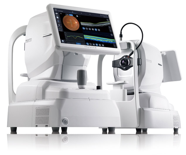
3D OCT & Fundus Camera, Totally integrated system combined with PC. Provides OCT and Fundus data on one Screen.
All-in-One HOCT is easy to use.
One button, it creates a High-speed Scan and a High-quality Image, giving greater insight for the ophthalmology clinic. One space, allows the acquisition of various types of analysis and diagnosis results.
Easy to use and results are outstanding and easily follow. Huvitz All-in-One HOCT will be the icon for leading a new era of Optical Coherence Tomography(OCT).
HIGH-SPEED and HIGH-QUALITY
Incredible speed of 68,000 A-scan/sec. :
More Realistic and Clearer image in high resolution
Provides High-speed Scan, High-quality Image by using Huvitz’s outstanding optical technology and innovative image software. Shows extensive information, such as 3D structure of Retina, Macula’s thickness and separation, in a vivid image.
High Resolution Image – min. 60 lines/mm of central Fundus
Creates 3μm OCT Digital Resolution medical images, allows more precise Retina observation and useful follow-up examinations.
Accurate and Stable Image Averaging
It is very important to obtain high-quality images that are accurate and stable in all OCTs. However, it’s not easy to capture these due to patient eye movement over the period of the test. The HOCT detects fast eye movements with image processing algorithms of fast Scan Speed* and Smart Viewing Technology (SVT)** and scans up to 68,000 points per second, and calibrates to create a high-quality optical image. HOCT can acquire high-quality images without any repetitive operation for first time users.
* 68,000 A-scan / sec., Less than 1.4 sec. in 6×6 mm2 3D shooting
** Smart Viewing Technology : Huvitz’s Speckle-Noise-Reduction System & Pre-Acquiring Algorithm to acquire high-quality images
Vividly Visualized Retinal Layers
Visualizing with precise B scans and smooth 3D images at faster scan speeds makes it easier to observe pathological shapes and status in stratified Retinal Layers.
It is also useful to further elucidate the pathological rheobase of Macula and Optic Disc, including factors that impair Photoreceptor Function, Retinal & Choroidal Vasculature (vascular system) in a slice image for Retinal Layer consists of 7 pieces.
Brightness Level Adjustment
Precisely identify lesions by minutely adjusting image’s brightness and contrast. In this way, specific parts of lesions can be highlighted which help users to easily see details.
ONE for ALL SYSTEM
3D OCT, Fundus Camera, Built-in PC :
Combined-One is more accurate and useful
By combining OCT, Full Color Fundus Camera, and PC, it can generate high resolution images providing multi-purpose functions for diagnosis.
It saves both time and space by performing frontal view(Enface) of eye diseases, Tomography, cross-compare and diagnosis in a single run.
Combined-One
Provides maximum psychological stability to the patient without re-shooting and reduces stress during shooting*.
Easily checking lesion’s position by Fundus Image, it precisely guides the location of the OCT Scan Image.
*Motion detection technology:Smart Scan Technology (SST) is applied to achieve perfect images without re-shooting even though there’re flicker or movement (see Smart Scan page).
Compact Design – it can be installed in a small space
Thanks to HOCT’s space-saving design its perfect for hospitals and research areas with many diagnosis devices and treatment equipment, It can maximize the convenience of users as well as patients, thus saving time and space.
Web Browsing System to view data anytime, anywhere
Patient’s test data can be analyzed anywhere on the Internet. You can check and analyze all data of HOCT through Web Browser such as Internet Explorer, Safari, Chrome without installing special software separately.
USER FRIENDLY
Auto Tracking & Auto Shooting :
makes it easy to use and obtain reliable data
HOCT is smart.- Obtains reliable data with minimum deviation of image quality according to user’s measurement proficiency.
By selecting the measurement mode it provides exact and more accurate images.
Fast and Stable Full Auto Mode
Simply press the button once to capture the image quickly and easily without any errors with Auto Tracking, Optimize, Auto Shooting at the correct position.
Depending on the application, select Semi Auto to obtain more detailed images.
Semi Auto Mode for more precise images
You can obtain a more precise image by shooting Semi Auto Mode turning one’s gaze to the side for patients with eye diseases such as cataract, strabismus, or optic disk and peripheral measurements. Semi Auto Mode can also be applied to eyes with weak signals.
XY alignment, focus is automatically adjusted, and manual operation during auto adjustment is also possible.
Focusing and Firing functions can be judged and involved by users so that users can obtain images in an intuitive way.
SMART SCAN
Start and finish instantly through only one-click :
Speedy process reduces errors in forward looking of patients
It provides more convenience and accuracy by offering an easy and various scanning function with Macula, Optic Disc, and Anterior.
Wide Area Scan(12mm x 9mm) for efficient diagnosis
A quick scan covers Macula and Optic Disk areas extensively.
By scanning around Optic Disc or Macula for patient’s pathological status, you can check the Thickness Maps between RNFL(Retinal Nerve Fiber Layer), GCL(Ganglion Cell Layer) and RPE Layers.
Smart Scan Technology with motion detection technology
Image analyzer with Huvitz’s unique Smart Scan Technology (SST) obtains a complete and perfect C-scan image by detecting any motion of eye flicker or movement that would prevent disappearance of scan line and image collection during measurement.
Providing various and useful scan patterns
12 different patterns make it available to choose and apply the optimized pattern to the main symptoms or the area of retinal disease without repetitive work or time-wasting.
ACCURATE ANALYSIS
Accurate segmentation and measurement :
Analyze pathology status from various perspectives.
A complete analysis helps you observe symptoms, illnesses and progress of each patient at a glance. Key indicator values compared to Normative Data are displayed in table and chart format.
Progression to track pathological changes
OCT scan and fundus image of a patient can be compared at a glance to sequential measurement results from baseline to present. Progression from past to present helps analyze disease progression and treatment process. Thickness, Enface, and ETDRS can be superimposed on the IR or Fundus at each measurement point so that the change in thickness of nerve fiber can be confirmed according to the transition. It also provides a trace graph so you can study at a glance.
Compare before and after patient’s symptom
You can compare and analyze the baseline data of a patient with the current data.
3D modeling in high speed and wide area
High-speed, wide area(12mm x 9mm) 3D images help you quickly and comprehensively understand the condition of the Retina. Also, layer thickness maps can be used from ILM to RPE, respectively and
Morphological changes on the measured surface of the layers can also be visually confirmed.
OU to cross-analyze function of binocular
Provides comparative analysis for Macular Thickness, RNFL Thickness, ONH(Optic Nerve Head) of binocular.
Summary : Monocular-Scan and OCT / Fundus image
Provides a summary analysis of Macula retina, RNFL, ONH at a glance. Helps identify whether follow-up examinations are needed or not. Easy to explain the results to the patient after diagnosis.
ANTERIOR MEASUREMENT
One Single System :
Start and finish in one place, making patient more comfortable.
Anterior Segment Module allows measurement and analysis of cornea thickness, angle and 3D image. It helps users work more efficiently by acquiring both anterior and posterior in one place.
9mm Wide Chamber View
Measurement of ACA (Anterior Chamber Angle) between cornea and Iris allows diagnosis and management of angle-closure glaucoma patients.
9mm High resolution Cornea Thickness Measurement
The 9mm high-resolution Cornea Scan provides an objective view of the structure of the eyeball and displays a cross-sectional image of the measured corneal thickness.
Corneal Thickness Map
Corneal’s irregularity, Thinnest point, etc. can be identified with a corneal thickness map to visualize the patient’s corneal thickness at a glance.
FULL COLOR FUNDUS IMAGE
Insight of Posterior Segment of Eye :
for Comprehensive diagnosis
Color Retinal Images optimized with high-resolution and contrast are very useful in analysis and clinical diagnosis. Best images are provided by Low intensity of flash, fast capture speed, quiet operation, small pupil mode and automatic flicker detection.
High resolution and performance 12 Megapixel Camera
High performance camera with Motion Artifact Suppression Technique provides high resolution images and also its low intensity of flash, fast & quiet operation maximize measurement quality.
Auto-Detection Of Pupil Size and Auto Flash Level Function
It accurately measures the pupil size and automatically adjusts the intensity of light according to pupil size. Even patients with small pupil size can be easily measured without switching mode. Selecting Small Pupil Mode, to be adjusted more intensive light for the small pupil size.
Panorama function for wide range of peripherals
Multiple built-in capture color fundus images at different positions and automatically stitch them to optimized total overview. By providing high-resolution images with minimal distortion, you can immediately see key information for a comprehensive assessment of patient’ eye.
Fixation Target for flexible configuration
Fixation target can be set on the display for fine adjustment of a specific part of the eyeball.


